With the sorely inadequate supply of donated human organs – from kidneys to livers and lungs to hearts – available in Israel, scientists here have invested years of research into producing artificial tissue for transplant. But until the technique can proceed, a way must be found to create effective arteries, veins and capillaries to supply blood to the artificial tissue.
Scientists from the Technion-Israel Institute of Technology in Haifa and the Weizmann Institute of Science in Rehovot in Israel have discovered the mechanical forces that influence the spatial organization of blood vessels. The Haifa scientists are experts in tissue engineering, while the Rehovot team are adept in the physics of complex systems. This combination of knowledge helped them to understand in detail how mechanical forces can direct the orientation of developing blood vessels. Their findings, which were published in the scientific journal Nano Letters, could advance methods of growing artificial tissue for transplant.
The present research was led by doctoral student Shira Landau and Prof. Shulamit Levenberg of the Technion’s Faculty of Biomedical Engineering and doctoral student Avraham Moriel and Prof. Eran Bouchbinder of the Weizmann Institute’s chemical and biological physics department, in collaboration with Dr. Ariel Livne, a former postdoctoral researcher at Weizmann’s department of molecular cell biology.
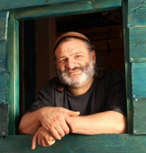
Levenberg, a 50-year-old modern Orthodox Jew and wife who has six children and lives with her family in the Galilee, has since 2015 been a full professor of biomedical engineering at the Technion. She began her studies completing a bachelor’s degree in biology from the Hebrew University of Jerusalem and went on for a Ph.D. in molecular biology at Weizmann. She joined the Technion after doing five years of post-doctoral work at the prestigious Massachusetts Institute of Technology. There, she built biological scaffolds to coax stem cells into developing into specific cell types.
Some 13 years ago, Levenberg was included in Scientific American 50 listing, selected by the prestigious science journal Scientific American, which honors people, teams, companies and organizations whose accomplishments in research, business or policy-making demonstrate technological leadership. Previous winners of this distinction include the two founders of Google and various Nobel Prize winners.
Cells, whether in the body or in lab-grown tissue, constantly interact with the extracellular matrix (ECM) – a very complex molecular network that, like a platform, provides structural support for cells. Until recently, scientists had assumed these interactions were primarily biochemical. But the researchers have now realized that mechanical interactions – for example, the ability of cells to sense various properties of the ECM and respond in kind – also play significant roles in cell development and function.
One of the scientific challenges to producing artificial biological tissues for transplant is that – like the real thing – they must contain a network of blood vessels to ensure a steady supply of oxygen and nutrients. Vital to the successful integration and survival of the implant is the directional order of this network – that is, the blood vessels must organize themselves in the same direction.
In Levenberg’s laboratory, she and her team developed a platform designed to improve tissue generation and self-organization for transplantation. The technology is based on three-dimensional scaffolds made of polymers, which are large molecules composed of many repeated sub-units.
Biological cells that are essential for the development of blood vessels are seeded on these polymeric scaffolds; studies have proven this technology viable and robust. In a series of studies, Levenberg and her team had previously used this platform to examine the mechanical sensitivity of vascular networks. In particular, they noted that mechanical forces have a strong influence on the properties of these networks, especially on the directions in which they grow and develop.
In 2016, Prof. Levenberg and Dr. Dekel Rosenfeld (then a doctoral student in her lab) showed how an original stretching system, which applied forces to the artificial tissue, allowing it to be stretched. This affected the cells’ biological processes including differentiation, shape, migration and organization in the structures – as well as the geometry of the emerging tissue, its maturity and stability. “We then wanted to understand how this process works and how to control it,” recalled. Landau.
In the new study, the researchers considered two types of stretching forces that can affect the development of blood vessels. These two, known as dynamic-cyclic stretching and static stretching, can lead to the emergence of directional order in networks of the blood vessels. The researchers discovered that the biophysical mechanism behind each of these two processes is basically different.
The study led to the establishment of a tensile stretching protocol – one that allows the controlled generation of optimal tissues, including stable, rich networks in which the blood vessels have a well-defined directional order. The researchers believe that these results and insights they provide will advance the possibility of engineering blood vessels in tissue with structures and directionality to enable their successful transplantation in patients.
In the past, researchers had managed to transplant only engineered muscle tissue with small blood vessels. But Levenberg’s previous medical breakthrough of engineering a muscle flap for repair of a large soft tissue defect – on which the Technion holds a patent – could eliminate the need for complex surgeries.
In tissue reconstruction, there are two techniques to deal with the clinical challenges involved in the successful restoration of tissue defects, Levenberg said. In one of them, there is a graft of tissue transplanted to the damaged area. The blood vessels in the body penetrate the tissue and nourish it with a blood supply. The second technique involves a flap transplanted to the damaged region together with its own blood supply.
Flaps are used for treating injured areas that do not have an adequate blood supply and when blood vessels do not develop following transplantation. This occurs when damaged soft tissues do not close or there is exposed bone, tendons or cartilage, Levenberg said.
The tissue was grown in the lab by Technion researchers and then implanted to the region around the femoral artery and veins in the thigh before being transferred as a flap.
The flap is the stage where tissues can be transferred with their own blood supply and are joined to the blood vessels in the region of transplant to repair large defects, such as in the abdominal wall region.
In 2007, Levenberg’s team created cardiac tissue in the lab from human embryonic stem cells and managed to bring about the creation of tiny blood vessels within the tissue, which makes possible their tissue’s implantation in a human heart. The culture was carried out in three dimensions on a scaffold made of self-destructing sponge material that the researchers also created in their lab. The technique is aimed eventually at helping patients who have cardiac insufficiency due to heart attacks.


